I. Introduction:
Squid are marine cephalopods of the order teuthida. Like all cephalopods (cephalopod means head-footed) squid have a distinct head, bilateral symmetry, a mantle and arms. Also all cephalopods have well developed eyes and brain, their head projects into a crown or group of flexible, muscled, arms, well developed mouth with jaws and a hard parrot-like beak for tearing off food pieces, they are all active and quick predators found only in marine habitats, and all cephalopods can swim quickly by expelling water from their mantle cavity. Also squid belong to the Squid are able to adapt to their surroundings even in the depths of the Mediterranean Sea and the Atlantic Ocean, which is where they mainly reside. Squid usually live only one year and measure up to 60 cm; although, the giant squid may live up to two years and measure up to 13 m. The squid’s taxonomy is the following, kingdom: Animalia, phylum: Mollusca, class: Cephalopoda, order: Teuthida, family: Loliginidae, genus: Loligo, species: Brevipinna.
The squid’s body is divided into the head, the neck, and body trunk. The main internal body is enclosed by the mantle, which has a swimming fin at each side. The skin of the squid is covered with cromatophores, cells that change pigment color in order to camouflage from prey and predators. Below the neck section of the body there are four pairs (eight) of arms, one pair of tentacles, and also, located in the center of the circle of arms, the mouth. The arms all have two rows of stalked suckers on the underside of them, these help to secure food. The arms serve to hold food in place while eating; in males the fourth arm is used to transfer spermatophores. The tentacles are used to grab food and pull it over to the arms. Around the mouth are the buccal and the peristomial membranes. In between these and inside the mouth is the horny beak used to tear off food pieces. In the neck section are located the eyes and the funnel or siphon. The squid’s eye is a very advanced eye, more similar to vertebrate ayes than to any invertebrate eye. This is an example of convergent evolution. The funnel is a muscular tube that extends out from the mantle collar. The funnel directs the water expelled out from the mantle cavity in order to control movement. Also wastes and ink are also expelled through the funnel. The mantle is made of muscular tissue and it protects the visceral mass and the mantle cavity. In the mantle are two swimming fins. Currents of water enter the mantle cavity through the mantle collar and are propelled as water jets through the funnel for propulsion.
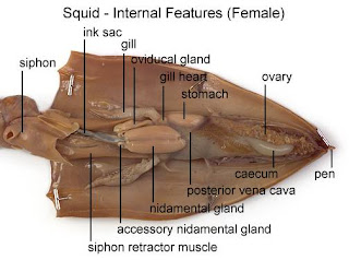
The internal anatomy of the squid is composed of many parts including organs and complete organ systems. From top to bottom are the pen, the cecum, the ovary, the posterior vena cava, the stomach, the gill heart, the nidamental gland, the gills, the oviductal gland, siphon retractor muscle, the ink sack, and the siphon. The pen is an endoskeleton composed of the dorsal, internal, chitinous Shell, and other elements constructed of chondroid, a clear, firm, cartilage-like material. The pen is what gives the mantle definite shape and structure. The cecum is a digestive organ directly connected to the stomach. The ovary, the dissected squid is a female, releases eggs into the mantle cavity. The posterior vena cava carry collected blood form the posterior body regions into the branchial hearts. The stomach is located next to the cecum, the ovary, the stomach and the posterior vena cava. The ink sac is located between the gills and just above the siphon or funnel.
The squid has a well developed circulatory system including a vast capillary system that reaches all body tissues. Also all the blood vessels are lined with epithelial tissue. The squid’s active predatory existence requires for a quick replenishment of nutrients, its circulatory system provides just this. The squid’s circulatory system consists of three separate hearts, the systemic heart and two gill hearts. The gill hearts receive the blood returning from the body tissues and pump it into the gills. This forces the blood into the thin capillaries of the gills, where the gas exchanges of carbon dioxide passing into the water and oxygen passing into the blood occur. The oxygenated blood then is passed to the systemic heart, where it is pumped to the body tissues via the arterial system.
When squid eat they tear off food pieces with their parrot-like beaks. One pair of salivary glands produces digestive enzymes and mucus, the other pair produces a poison. When a squid bites its prey the poison injected into the prey subdues it and makes it easier for the squid to eat. Every bite of food is mixed with saliva and is passed to the stomach through the esophagus. The stomach is a muscular organ and it kneads the food with digestive secretions. After the food is mixed and kneaded it goes on to the cecal sac where digestible food particles are sorted from non-digestible one, and then the digestible food particles are absorbed through the cecal walls, and non-digestible waste is expelled through the anus and out the funnel. The digestive modifications found in the squid are adaptations for rapid digestion of food that supports the squids’ active carnivore existence.
The squid has two compact, sac-like kidneys, and that sac also includes the pancreas. Through the middle of each kidney is a large systematic vein that leads from each kidney to one of the gills. Between the kidney and the vein is where the waste is transferred from the blood. The waste is mostly ammonia and because squid have no bladder and waste expelled through the mantle funnel.
Squid have one of the most highly developed nervous systems of all invertebrates. The squid brain is the largest invertebrate brain and is composed of the typical molluscan ganglia that have been fused and concentrated together to form a rather large brain mass. The brain is divided into various ganglia regions that surround the esophagus. These ganglia regions are so well divided that scientists have been able to prove some actions correspondent to some regions. The most important ganglions are the brachial and the visceral, which receive nerve inputs from the arms and tentacles, and receive and processes input from body organs. Extensive studies on squid have demonstrated that they have a great learning ability, which is credited to their sophisticated brain. The brain is protected with a cartilage “skull” or cartilage.
Male squid have a sac-like testis that produces sperm. Female squid have a single ovary that releases eggs that travel through the oviduct to an oviductal gland. Here the eggs are covered with a gelatinous covering as they are released for fertilization. During mating the male uses a specialized arm to pluck a spermatophore from its reservoir and inserts it into the female’s opening. The spermatophore wall disintegrates releasing the sperm into the female mantle cavity. The eggs leave the mantle cavity and the female grasps them with its arms and are fertilized by sperm exiting the female’s funnel. The female attaches the eggs to a sea rock and once these touch the sea water a gel-coating hardens to help protect the eggs.
II. Purpose:
The purpose of the laboratory was to get us familiarized with the internal anatomy of some animals in order to go back and look at the human anatomy. Also to be able to describe the organism in specific terms, discuss the characteristics of the organism’s taxon, and the symmetry of the organism and peculiarities that the specific animal has.
III. Materials:
1. Dissection Kit
2. Dissection Tray
3. Gloves
4. Apron
5. Security Goggles
6. Preserved Squid
7. Biology Text or Another Reference
8. Plastic Bags
9. Notebook
10. Dissection Pins
11. Labels
IV. Procedure:
1. Pin the squid to the dissection tray in the dorsal position.
2. Identify the external anatomy: arms, the mantle the eyes, the tentacles, the siphon or funnel and the swimming fins located on the mantle. Also spread the arms in order to locate and observe the buccal mass.
3. Flip the squid in order to look at the extension of the pen,
4. Flip the squid back to the posterior view of the mantle and make an incision up the middle using a pair of dissection scissors with one blunt tip in order to prevent cutting any internal organs. Use the dissection tray to pin the two mantle flaps on either side of the visceral mass.
5. Locate al the internal organs and organ systems which should all be clearly visible.
martes, 7 de octubre de 2008
lunes, 6 de octubre de 2008
VI. Discussion:
In preparation for the dissection, a great deal of information was looked up in order to know what the steps to go through with the dissection were. After all the information was gathered, the end result was a compilation including a step by step process to dissect a squid. The animal that was dissected was a squid so everything worked out great without any major problems. The information that was used was very useful and precise in stating the easiest and most effective way of dissecting a squid.
During the process of the dissection there were some minor problems; nevertheless, the lab was completed without much strain. One of these problems was that the squid was very dehydrated and didn’t have a squid’s usual form; it was bent in an unusual way. This was a problem to begin with because when the mantle was being cut, pieces of the pen started to show up. Then after that side of the mantle was completely cut and the pen was removed, it was observed that the side that had been cut was the dorsal instead of the ventral. After this, the ventral side of the squid was cut open and it was then possible to locate the organs that were shown in many f the diagrams of the information found for the dissection.
VII. Conclusion:
This laboratory had quite a few of purposes, of which includes learning the organism’s symmetry and some peculiarities of the organism. In the laboratory it was possible to identify specific parts and understanding the use of these. Properly using the dissection kit and the lab materials it was possible to effectively dissect the cephalopod and identify the majority of its parts.
I had never dissected a cephalopod before; the only organism I had dissected was a crayfish last year. Even though I had dissected another organism before this experience was new because it was a whole new specimen and it was a different procedure. The internal and external anatomies were successfully identified, including their each specific function.
VIII. Reflection:
I really liked this laboratory and I worked hard on it. I am very interested in biology so I was impressed by what I saw right after I opened the mantle cavity. I was intrigued by the anatomy of the squid and so I was very anxious to locate and identify everything in the squid. I got a little frustrated when I cut the wrong side of the mantle but was relieved when I realized that it didn’t really matter. I really liked the activity and would hope if we got around to doing some more dissections apart from the final exam in May.
IX. References:
1. Fox, R (2001). Invertebrate Anatomy OnLine . Retrieved October 5, 2008, from Lolliguncula Web site: http://webs.lander.edu/rsfox/invertebrates/lolliguncula.html
2. Herreid, C (1999, 11, 15). Evolutionary Biology. Retrieved October 5, 2008, from Laboratory Tutorial Web site: http://www.bio200.buffalo.edu/labs/tutor/Squid/
3. Squid Dissection: From Pen to Ink. Retrieved October 5, 2008, Web site: http://www.nhm.org/seamobile/PDF/clasacts/sqd%20i.pdf
4. Squid Dissection. Retrieved October 5, 2008, from Squid Dissection Web site: http://www.biologycorner.com/worksheets/squid.htm
5. squid. (2008). In Encyclopædia Britannica. Retrieved October 07, 2008, from Encyclopædia Britannica Online: http://www.britannica.com/EBchecked/topic/561782/squid
Suscribirse a:
Comentarios (Atom)


